Usefulness of the lateral arm free flap for hand defect reconstruction
Article information

Abstract
Purpose
Although various surgical reconstruction methods are available for multidigit defects or soft tissue defects of the hand, they often require a thin flap due to the hand’s unique physical characteristics. We performed a total of 13 cases of lateral arm free flap surgery at our hospital. The purpose of this study was to report the usefulness of this technique.
Methods
This study included the following cases: four cases of multiple digit amputations, seven cases of hand dorsum skin defects, one case of first web reconstruction due to web contracture, and one case of an index finger bone and soft tissue defect with only the ulnar-side neuro-vascular bundle remaining. After emergency simple debridement, subsequent reconstruction was performed using a lateral arm free flap.
Results
All 13 cases healed without necrosis. Patients who underwent finger amputation were able to preserve the length of their fingers without any additional amputation. Patients who initially had hand dorsum defects were able to maintain grasping function after flap stabilization. The patient with web contracture was able to recover the lost pinching function, and the patient with a combined loss of osteoarticular and soft tissue of the index finger maintained the shape and length of the finger despite the absence of joint function.
Conclusion
The lateral arm free flap is a useful reconstruction method that can be easily performed with a single session of regional anesthesia. Since the flap is thin, it is suitable for hand defect reconstruction. If necessary, vascularized bone grafting is possible.
Introduction
Various surgical methods are available for reconstructing multidigit defects or large soft tissue defects of the hand. Even though there are many types of flaps, either pedicled or free flaps, the advantages of free flaps over distant pedicled flaps in hand reconstruction are well documented. Free tissue transfer typically requires only one operation and allows all other reconstructive procedures to be performed at the same time. Additionally, rehabilitation therapy can be initiated as soon as the patient recovers from anesthesia, and postoperative edema and stiffness are reduced. These benefits are not possible with the use of pedicled flaps [1-3].
To restore the large defects of hand dorsum, a thin skin with minimal subcutaneous tissue layer maintaining sensation is necessary. There are several types of free flaps, including lateral arm free flap (LAF) and thoracodorsal artery perforator flap (TDAP), that can be used for this purpose. At our hospital, we have chosen to use LAF, which can be performed with a single regional block (brachial plexus block) and can also be used as an osteocutaneous flap when needed. Traditionally, LAF has been used in various reconstructive surgery, including the head and neck surgery, and we would like to report on the usefulness of LAF focusing on the hand reconstructive surgery.
Methods
This study was approved by the Institution Review Board of Duson Hospital (No. 2022-10-013), and the patients provided informed consent for the publication of this report including all clinical images.
We conducted a retrospective study of 13 cases of LAF surgeries performed at our hospital between August 2015 and July 2022. The patient group consisted of 12 males and one female, aged between 24 and 60 years, with an average age of 44 years. The surgeries were performed by three surgeons for multiple digit amputations (four cases), soft tissue defects in the hand dorsum (seven cases), the first web space contracture (one case), and osteocutaneous defects (one case). The time from injury to flap surgery ranged from 3 to 23 days, with an average of 11 days.
The flap was harvested in a hemostatic operative field using a tourniquet, based on the predetermined size and design. The central axis of the flap was defined as the line connecting the deltoid insertion to the lateral humeral epicondyle. Once the flap was outlined, the dissection proceeded from the proximal to the distal direction. The incision was made posteriorly to the lateral intermuscular septum over the lateral head of the triceps muscle. The deep fascia was incised and elevated, and the septocutaneous perforator artery passing through the intermuscular septum is confirmed. To prevent the skin and fatty layer from separating, three stitches were used for suturing, and after leaving the sutures long and cutting them, we continued to elevate the flap in the anterior direction. The distal fascia dissection was carried out up to the humeral periosteum, and we marked the lateral antebrachial cutaneous nerve during dissection. The posterior radial collateral artery (PRCA) was ligated at the branching point to the anterior radial collateral artery. In cases where the humerus bone was harvested together with the flap, the intermuscular septum connected to the periosteum was harvested simultaneously, as it often gives rise to one or two branches from the PRCA towards the bone.
The artery of the harvested flap was anastomosed to the dorsal branch of the radial artery using an end-to-side technique, or to the common digital artery using an end-to-end technique. One to two venous anastomoses were performed using vena comitans and subcutaneous vein. In cases where primary closure of the donor site was not feasible, a subsequent full thickness skin graft (FTSG) was performed. In cases of multiple digit amputations, flap division and inset procedure was performed at 3 weeks, once flap circulation stabilized. When dividing the flap, z-plasty technique was used to facilitate the flap inset.
Results
All 13 cases healed without signs of necrosis. The widths of the harvested flaps ranged from 4 to 7 cm (mean, 5.62 cm), and the lengths ranged from 8 to 14 cm (mean, 10.85 cm). In two cases, primary closure of the donor sites was not possible, so subsequent FTSGs were performed. There was no observed limitation of movement in the elbow joint. However, in one case, diminished sensation and discomfort in the forearm were reported (Table 1).

Patients' data
Patients with digit amputations were able to preserve the length of their fingers without further bone resection. Through rehabilitation, patients who initially had hand dorsum defects were able to maintain the grasping function of the hand. The patient with web contracture was able to recover the pinching function. And the patient with simultaneous loss of osteoarticular soft tissue of the index finger was able to maintain the shape and length of the finger despite having no joint function.
1. Case 1
A 57-year-old male patient was admitted after sustaining hand injuries from a press machine. He had multiple crushing injuries and amputations at the base of the middle phalanx of the index finger and at the level of the proximal phalanx of the long finger, ring finger, and small finger. A 6×13-cm flap was harvested and used for surgery. The arterial anastomosis was performed on the third common palmar digital artery, and the venous anastomosis was performed on the cutaneous veins. Three weeks after the surgery, flap division and inset procedure was performed on the third web space, followed by division of the remaining fingers a week later (Fig. 1).

Case 1. (A) Multidigit amputation state due to a presser machine. (B) X-ray image after trauma. (C) Intraoperative photograph of flap harvest. (D) Postoperative photograph of the recipient site. (E) After the first release of the third web space. (F) Postoperative photograph of the recipient site after 6 months.
2. Case 2
The 56-year-old male patient was admitted after sustaining injuries to his hand from a thresher. He had comminuted fractures in the second and third metacarpals of the right hand, as well as multiple thermal injuries and skin defects on the dorsal surface of the hand and volar side of the forearm. Initially, surgery was performed to address the fractures, and an open dressing was applied to the skin defects. To prevent further skin necrosis, debridement and flap surgery were performed using a 5×10-cm flap that was harvested. The arterial anastomosis was performed on the radial artery dorsal branch, and venous anastomosis was performed on the adjacent cutaneous veins (Fig. 2).
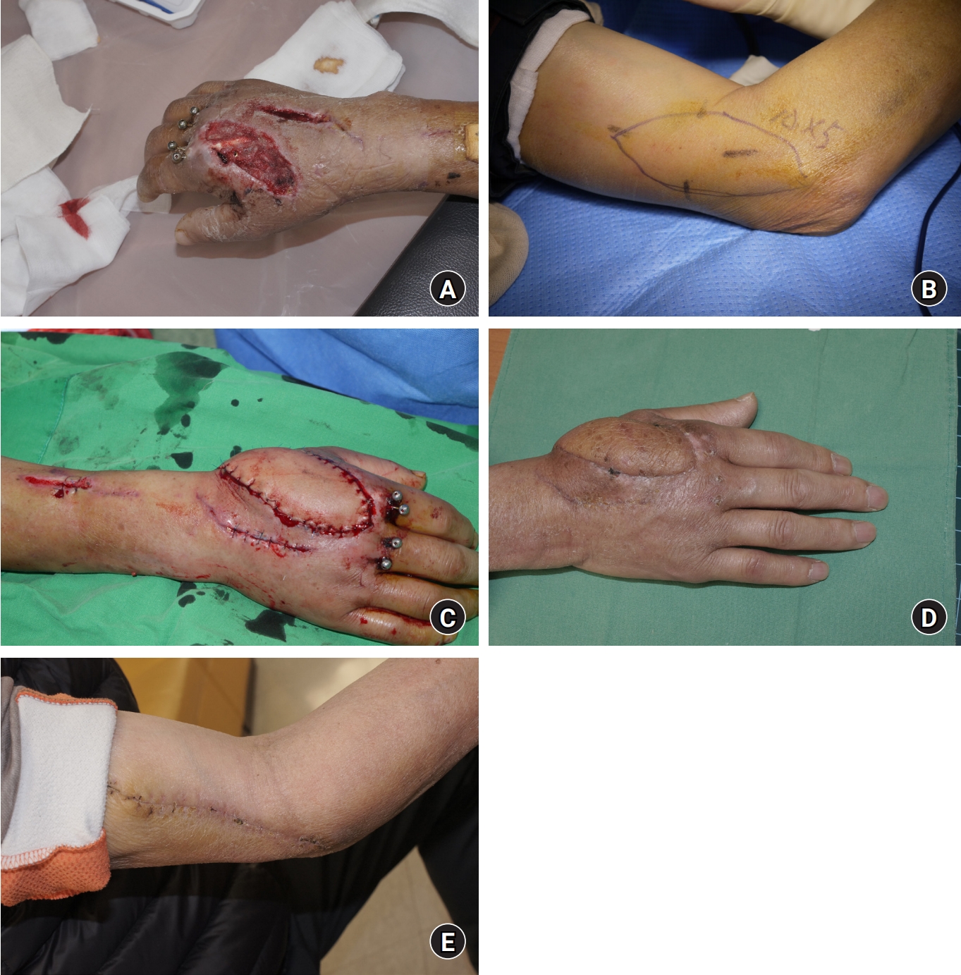
Case 2. (A) After primary debridement of necrotic tissue in the right hand. (B) Preoperative design of the lateral arm free flap on the right upper arm. (C) Postoperative photograph of the recipient site. (D) Postoperative photograph of the recipient site after 3 months. (E) Postoperative photograph of the donor site after 2 months.
3. Case 3
A 38-year-old male patient was admitted due to a crush injury and incomplete amputation injury of the index finger. He had severe skin and tissue crush injury at the radial side of the midportion of the index finger and also had severe bone loss in the middle and proximal phalangeal bones. Initially, debridement, wound closure, and fracture fixation were performed, but skin necrosis progressed gradually. To address the skin and bone defects, a free osteocutaneous lateral arm flap was performed. A 6×8-cm skin paddle and a 1×3-cm segment of the proximal humerus were harvested. The arterial anastomosis was performed on the radial digital artery of the index finger. The bone fragments were aligned and stabilized using metal plates and Kirschner wires (K-wires). Five months after the surgery, he was able to maintain his index finger close to normal shape, and he was able to grab objects by flexing the metacarpophalangeal joint (Fig. 3).

Case 3. (A) Initial X-ray image after trauma. (B) Initial photograph after trauma. (C) Necrosis of the soft tissue on the radial side of the index finger after primary surgery. (D) Intraoperative photograph after harvest of an osteocutaneous lateral arm free flap. (E) Postoperative photograph of the recipient site. (F) Postoperative X-ray image of the recipient site. (G) Postoperative photograph of the recipient site after 5 months (dorsal aspect). (H) Postoperative photograph of the recipient site after 5 months (volar aspect).
4. Case 4
A 40-year-old male patient was admitted with a degloving injury to the palm of his hand inflicted by getting caught in machinery. He had concomitant fractures of the proximal phalanx of the long finger and ring finger, for which a fracture fixation with K-wires and wound closure were performed while repairing two digital veins. Due to extensive necrosis of the palm skin including the first web space, a decision was made to perform a LAF. A 5×14 cm long flap was harvested by extending it to the forearm. The arterial anastomosis was performed on the radial artery dorsal branch and two cutaneous veins were used for the venous anastomosis to complete the surgery (Fig. 4).

Case 4. (A) Initial photograph after trauma. (B) Necrosis of the first web space and volar tissue of the hand after debridement. (C) Intraoperative photograph before harvest of the extended lateral arm free flap. (D) Postoperative photograph of the recipient site (dorsal aspect). (E) Postoperative photograph of the recipient site (volar aspect). (F) Postoperative photograph of the recipient site after 9 months.
Discussion
Various flap surgeries can be attempted to reconstruct hand injuries with multiple digit amputations and extensive skin defects. Anterolateral thigh flap (ALT), TDAP, radial artery superficial palmar branch free flap (RASP), and medial sural artery perforator flap (MSAP) are possible options, but ALT generally has a thicker flap, TDAP requires general anesthesia as a necessity, RASP cannot exceed 4 cm in width, and MSAP also requires additional general or regional anesthesia for surgery [4]. For debulking the thick flap like ALT, microdissection techniques can also be attempted, but it has the disadvantage of requiring additional surgical time.
An ideal fasciocutaneous free flap should meet the following criteria: substantial skin availability, a long vascular pedicle, thin and innervated skin, easy surgical access, and minimal donor-site defect [2]. The LAF is a fasciocutaneous flap with constant vascular anatomy, and since it contains a nonessential vessel, the vascularity of the limb is never compromised after harvesting the flap. The flap also has good venous drainage, requiring only one anastomosis. The skin around the elbow is very thin, ranging from 0.2 to 0.5 cm. Getting closer to the shoulder it becomes thicker, ranging from 0.5 to 1 cm. Therefore, if a thin flap is needed, the flap’s design should be placed around the lateral elbow area, while if a thick flap is needed, the flap should be positioned more toward the center of the humerus to obtain the desired thickness of the flap. If the flap is too thick, only the muscle and fascia can be transplanted by dissecting the skin and subcutaneous tissue of the flap, or a flap with skin grafted over myofascial tissue can also be used [2,5-8].
In addition, LAF can be performed under a single regional anesthesia with brachial plexus block, which is deemed safer than general anesthesia. It does not cause any significant complications at the donor site and allows surgery without changing the patient’s position in a single operative field. It is suitable for hand injuries as it can collect relatively thin skin flaps, and sensory recovery can be expected as a neurosensory flap that includes sensory nerves [5]. It can also be utilized as an osteocutaneous flap since a portion of the humerus bone can be harvested together [5,9].
The disadvantages of LAF include limitations in size and the potential for hair growth, among others. However, the limitation of length can be overcome by extending the LAF. When needed, a thin flap including sensory nerves can be harvested by extending it to the forearm. Complications and morbidity associated with LAF include lateral epicondylar pain and hypesthesia of the proximal lateral forearm skin, among others, which have been reported [10].
The limitation of the current study is that the number of cases is relatively small, and a direct comparison with surgeries performed using other types of flaps and their outcomes was not possible.
Conclusion
The LAF is a useful reconstruction method that can be easily performed under regional anesthesia with brachial plexus block. In addition, since the flap is thin, it is suitable for hand defect reconstruction. If necessary, vascularized bone-grafting can be performed by harvesting bone, and the sensate flap is also possible when the cutaneous nerve is harvested together. Therefore, it is a useful surgical method that can be widely used for hand reconstruction surgery.
Notes
The authors have nothing to disclose.
Funding
None.