Reconstruction of a soft tissue defect in the toe using a serratus anterior fascia free flap: a case report
Article information
Abstract
Toe injuries frequently occur as traumatic or oncologic defects. Compared with finger reconstruction, toe reconstruction has been rarely reported in the literature, because of the difficulty of toes’ relatively thin soft tissue envelope, their requirement for relatively challenging surgical techniques, and the slight improvements in gait. Therefore, toe reconstruction can be challenging for plastic surgeons, especially in cases with exposure of a tendon, bone, or joint. Herein, we present a case study of a 54-year-old woman with squamous cell carcinoma in situ (Bowen disease) that subsequently resulted in a defect on her second toe. A serratus anterior fascia free flap could be a good option for toe reconstruction due to its large caliber, lengthy pedicle, relatively easy dissection, and thin muscle bulk. We present our unique experience using the serratus anterior fascia free flap for the reconstruction of an oncologic toe defect.
Introduction
Although not as common as finger injuries, toe injuries also frequently occur as traumatic or oncologic defects. Compared with finger reconstruction, toe reconstruction is rare because of the difficulty of its relatively thinner soft tissue envelope, its relatively high demand for surgical techniques, and little gain in gait [1]. Nonetheless, toe reconstruction may still be beneficial if there is a strong individual desire for aesthetical restoration. Reconstructive microsurgeons are usually more familiar with toes as the donor sites in toe-to-hand, toe pulp, and few reports have documented the reconstruction of toe defects. So, toe reconstruction can be challenging for plastic surgeons, especially with exposure of a tendon, bone, or joint [1,2]. In the context of traumatic or oncologic toe defects, the surrounding tissue is often insufficient for reconstruction, leaving free flap as the only viable option. We consider that the serratus anterior fascia free flap offers ideal characteristics for toe reconstruction due to its large caliber and lengthy pedicle, relatively easy dissection, and thin muscle bulk [3,4]. We present our experience using the serratus anterior fascia free flap for reconstruction of oncologic toe defect.
Case report
This study was approved by the Institutional Review Board of Hanyang University Medical Center (IRB No. 2023-05-058). Written informed consent was obtained from the patient for the publication of this report including all clinical images.
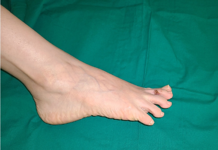
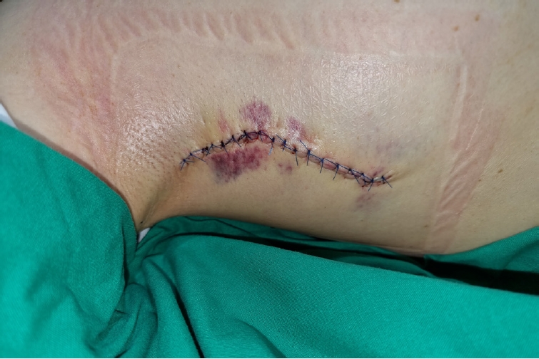
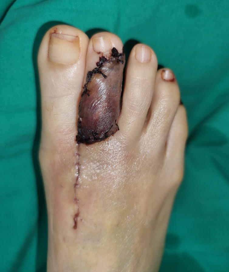
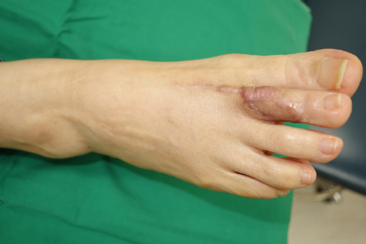
A 54-year-old female patient presented to the dermatologist with a lesion on the lateral side of her right second toe. A punch biopsy was performed, revealing a diagnosis of squamous cell carcinoma in situ (Bowen disease). The patient was referred to our plastic surgery team for surgical management. For the lesion of about 2.5×1.5 cm2 (Fig. 1), we performed a wide excision considering the safety margin and decided to perform a serratus anterior fascia free flap for the defect with bone exposure. A wide excision was made by setting the resection margin to about 5 to 8 mm, and the size of the resulting defect was approximately 3.5×2.0 cm2 (Fig. 2A). In order to dissect the first dorsal metatarsal vessel as the recipient vessel for the free flap, an additional incision of about 5 cm was made near the fist web space. While maintaining the supine position without position change, an incision was made along the right mid-axillary line to detach the subcutaneous tissue. After confirming the serratus anterior muscle below, the perforator artery was explored. After identifying a perforator that was relatively viable and whose pulsation was reliable as a pedicle, the fascia flap of about 4.0×2.5 cm2 and a pedicle of about 6 cm were harvested (Fig. 2B). The pedicle artery of the fascia flap was anastomosed to the first dorsal metatarsal artery in an end-to-end fashion, while the vena comitans was anastomosed to the vena comitans of the first dorsal metatarsal artery in an end-to-end fashion. After applying acellular dermal matrix on the muscle flap, split-thickness skin graft (STSG) was performed according to the size. The total operation time was 120 minutes, and the free fascia flap demonstrated relatively good matching with the surrounding tissues in terms of color, shape, and texture (Fig. 2C). The donor site in the right axillary area was closed primarily without tension (Fig. 3). The patient received dressing and conservative therapy for 2 weeks after surgery and was discharged for outpatient follow-up. One month after the free flap surgery, during outpatient follow-up, it was observed that the fascia flap engrafted well overall, but some portions of the skin graft did not take (Fig. 4). As the fascia layer was well-preserved, it was decided that it would be advantageous to remove the non-surviving STSG and perform full-thickness skin graft under local anesthesia. Three months after the surgery, it can be seen that the flap was engrafted well overall. Also, toe defect was all covered without complications such as infection, flap necrosis, and cancer recurrence (Fig. 5).

A 54-year-old woman with a squamous cell carcinoma in situ (Bowen disease) measuring about 2.5×1.5 cm2 on the lateral side of her right second toe.

(A) Toe defect was approximately 3.5×2.0 cm2 after wide excision of cancer. (B) A serratus anterior fascia flap of about 4.0×2.5 cm2 and a pedicle of roughly 6 cm was harvested. (C) After anastomosis, acellular dermal matrix was applied to the muscle flap, and then a split-thickness skin graft was performed. The fascia free flap demonstrated relatively good matching with the surrounding tissues in terms of color, shape, and texture.

The donor site in the right axillary area was closed primarily without tension. The linear incision was about 11 cm.

The fascia flap engrafted well overall, but the skin graft from about one-third of the proximal portion did not take well.
Discussion
Toe reconstruction remains challenging for plastic surgeons, especially in cases of traumatic or oncologic defects that require free flap due to insufficient surrounding tissue for reconstruction [1,2]. For this reason, amputations are much more common than salvage procedures. However, toe reconstruction, rather than amputation, provides superior functional and aesthetic results [5]. The primary consideration for selecting a free flap for toe reconstruction is its thinness. Thin free flaps such as superficial circumflex iliac artery perforator (SCIP) and thoracodorsal artery perforator (TDAP) flap have been reported in the literature to be suitable for toe reconstruction [5-7]. Among the thin free flap options, serratus anterior fascia also could be a good option due to its large caliber and lengthy pedicle, relatively easy dissection, and thin muscle bulk [3].
Firstly, the pedicle can be harvested wide and long, which is important for performing the anastomosis outside the defect, preferably without the need for a vein graft. The long and wide pedicle enables an anastomosis site to be located more proximally, allowing for easier and more stable microsurgery. Also, this allows for a reliable blood supply to the flap, reducing the risk of flap failure. In addition, the long pedicle allows for greater flexibility in positioning the flap, which can be crucial for achieving optimal coverage of the defect. Secondly, the serratus anterior fascia free flap has a relatively thin muscle bulk, which can be harvested thinner than SCIP or TDAP flap [6,7]. This is particularly important for toe reconstruction, where the soft tissue envelope is relatively thin. A thin flap also reduces the risk of bulkiness, which can lead to functional impairment or poor aesthetic outcomes. Therefore, the need for additional debulking surgery is reduced and reconstruction can be completed as a one-stage procedure. Thirdly, the dissection of the serratus anterior fascia free flap is relatively easy. The muscle is easily accessible and has a clear anatomical location, making it a reliable option for free flap reconstruction. Although there was only one case in this study, the total surgical time including toe tumor excision, flap harvest, and insetting was 120 minutes.
However, the serratus anterior fascia free flap also has some disadvantages. One disadvantage is that an additional skin graft, either split-thickness or full-thickness, is required to cover the raw surface of the fascia flap. In this case report, a STSG was performed initially, but a full-thickness skin graft was required to cover the non-surviving portions of the graft. This increases the complexity of the surgical procedure and can also lead to additional scarring. Another disadvantage is that accurate flap monitoring can be difficult due to the skin graft overlying the fascia flap. As a way to overcome these limitations, fluorescent indocyanine green (ICG) angiography can be considered. Evaluation of free flap perfusion is still based on subjective clinical features such as color, refill time, turgor, warmth, etc. In particular, color and refill time are factors that are difficult to observe in fascia flaps covered with skin grafts, making flap monitoring difficult. However, there is a research result that fluorescent ICG angiography is objective and effective for flap monitor [8], so it is thought that using ICG angiography for fascia flap monitor will be helpful. Although this report covers a single case of using the serratus anterior fascia free flap for soft tissue defects in the toes and dorsum of the foot, the number of such cases is currently increasing in our hospital. As the data accumulates to a sufficient level, we plan to conduct comparative studies with other reconstruction methods in the future.
In conclusion, the serratus anterior fascia free flap is a viable option for the reconstruction of toe defects, offering advantages such as a lengthy pedicle, thin muscle bulk, and relatively easy dissection. However, its disadvantages include the need for additional skin grafting and difficulties in flap monitoring. Overall, the decision to use the serratus anterior fascia free flap should be based on the patient's individual needs and the surgeon’s experience and preference.
Notes
The authors have nothing to disclose.
Funding
None.

