Clinicopathologic features of epidermoid cysts in the upper and lower extremities, including a case of malignant transformation in the palmoplantar region
Article information

Abstract
Purpose
Epidermoid cysts are common benign skin neoplasms derived from the pilosebaceous apparatus that usually develop in hair-bearing regions such as the head and neck. Epidermal cysts rarely occur in the extremities, especially in the palmoplantar region. Therefore, they can be easily misdiagnosed as warts or calluses. Here, we present our experience treating epidermal cysts in the extremities, including a very rare case of malignant transformation into squamous cell carcinoma.
Methods
This retrospective study enrolled all patients who underwent excision of epidermoid cysts in the upper and lower extremities from March 2006 to April 2021.
Results
Among 249 patients, there were 10 (seven male and three female patients) who had epidermal cysts in the extremities (4.0%). All four plantar cysts were located in weight-bearing areas. One palmar epidermal cyst occurred 33 years after the trauma. There was one case of a highly recurrent epidermal cyst on the heel that was eventually diagnosed as squamous cell carcinoma arising from the cyst lining on excisional biopsy. After wide excision with a margin of 2 cm, the resulting defect was reconstructed using a free thoracodorsal artery perforator flap.
Conclusion
Epidermoid cysts in the extremities, especially the palmoplantar region, are rare. Detailed history taking, including underlying diseases and trauma history, is helpful for diagnosis. Complete excision is necessary to avoid relapse and to confirm the final diagnosis, especially in cases suspected of malignant transformation arising from epidermoid cysts.
Introduction
Epidermoid cysts are common benign skin neoplasms that typically develop in adults. These lesions generally present as round nodules with a central punctum on the overlying skin [1] and are predominantly found in males vs. females (2:1) [2]. In general, these lesions remain asymptomatic until rupture, inflammation, or infection occurs. Histologically, they are lined with stratified squamous epithelium and filled with cheese-like keratin in the laminated layers. Most cases are derived from inflammation of the occluded pilosebaceous apparatus. Although epidermal cysts can appear anywhere in the body, they primarily develop in the hair-bearing regions, such as the head, neck, or trunk and fewer than 10% occur in the extremities [3]. Although most lesions are sporadic, multiple epidermoid cysts at atypical sites such as limbs may be signs of familial syndromes such as Gardner syndrome [4].
Epidermoid cysts can also be found in the palmoplantar regions which lack hair follicles [5-7]. The pathophysiological mechanisms of these palmoplantar epidermal cysts remain unclear. Previous studies have suggested epidermal implantation into the dermis following trauma or surgery and human papillomavirus (HPV) infection of the eccrine ducts as underlying etiologies [8-12]. Due to the relatively low incidence of glabrous skin, epidermal cysts may be confused with warts, calluses, ganglionic or synovial cysts, lipomas, or fibromas [2].
Furthermore, epidermal cysts rarely undergo malignant transformation into squamous cell carcinomas (SCCs; found in the head and neck, trunk, and testicle) [13-15]. Nevertheless, surgical excision is recommended to perform a final histopathological examination, especially for epidermoid cysts with the involvement of multiple and unusual sites. Complete removal of the capsule is recommended to prevent relapse. In this retrospective study, we share our experience of treating epidermal cysts found in the upper and lower extremities, including a rare case of malignant transformation to SCC on the heel.
Methods
Ethics statement: The study was performed in accordance with the Declaration of Helsinki after receiving approval from the Institutional Review Board of Seoul National University Hospital (No. H-2209-064-1357). Written informed consent was not required due to its retrospective nature, and the consent for publication of the clinical images was obtained from the patient.
The medical records of all patients who underwent surgical excision for epidermoid cysts located in the upper and lower extremities, between March 2006 and April 2021 in Department of Plastic and Reconstructive Surgery were retrospectively reviewed. Data on patient demographics, underlying disease, history of other operations, diagnosis, onset, operative findings, and histopathological findings were collected. The frequency of the epidermoid cysts found in the extremities was expressed as a percentage divided by the total number of epidermoid cysts on the whole body.
Results
Among 249 patients who underwent excision for epidermoid cysts, 10 patients (4.0%) presenting with lesions in the upper and lower extremities were enrolled in this study. Patient clinical information is summarized in Table 1. There were six out of 10 patients (seven male and three female patients) with epidermoid cysts in the palmoplantar region (1, palm; 1, finger; 3, ball area; and 1, heel), and four in the non-palmoplantar region (1, antecubital fossa; 1, posterior forearm; 1, posterior thigh; and 1, anterior tibial area). The epidermoid cysts were slow-growing masses varying in size from 7×5×3 mm to 45×23×16 mm. Plantar epidermal cysts in weight-bearing areas cause discomfort with ambulation, which means that such patients visit clinics relatively earlier than others. One patient presented with a palmar epidermal cyst that developed 33 years after a traumatic injury at the same site (case 1) (Fig. 1). Rupture of the epidermal cyst with foul odor and ulceration occurred in one patient (case 5) (Fig. 2). Three cases in the palmoplantar region were recurrent after previous treatment at other clinics (cases 1, 2, and 5). Considering the patient’s age in case 6, preoperative ultrasonography was performed for the differential diagnosis. Ultrasonography revealed a 1 cm-sized well-defined avascular cystic lesion with heterogeneous hypoechogenicity (Fig. 3).

Summary of cases

A 55-year-old male patient with a painful palmar epidermoid cyst that developed 33 years after penetrating trauma (case 1). (A) Preoperative and (B) intraoperative images showing the gross specimen.
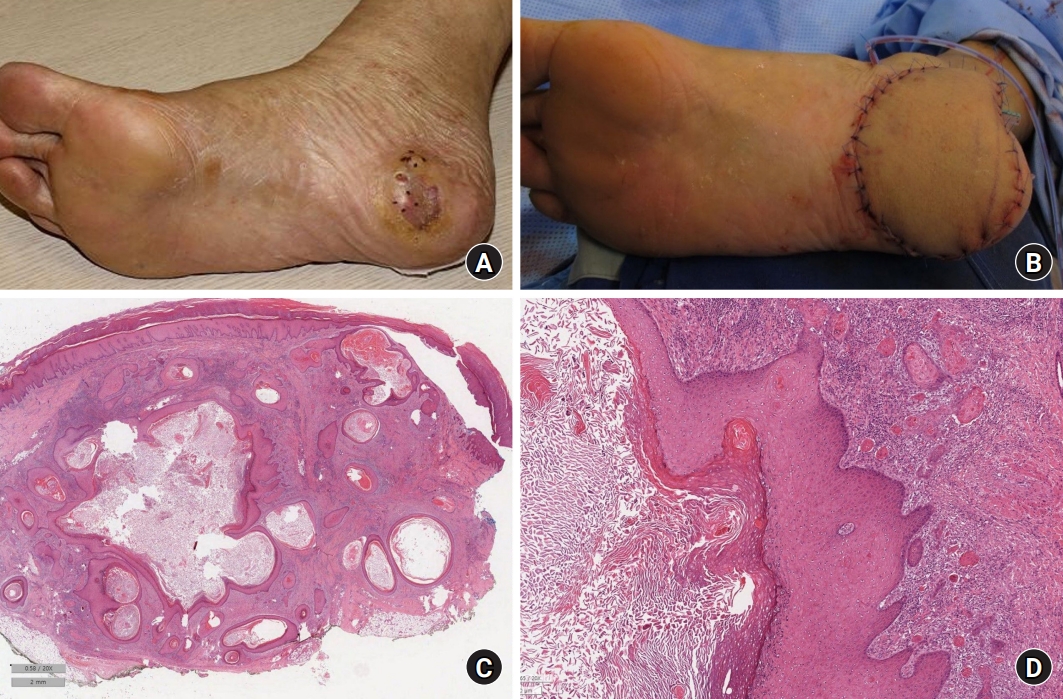
A 46-year-old male patient with a recurrent plantar epidermoid cyst (case 5). (A) Preoperative image showing the epidermoid cyst with overlying skin changes. (B) Postoperative image after wide excision and reconstruction with a free thoracodorsal artery perforator flap. (C, D) Microscopic images showing malignant transformation into squamous cell carcinoma arising from the cyst wall (H&E stain; scale bars, 2 mm in panel C and 200 μm in panel D).
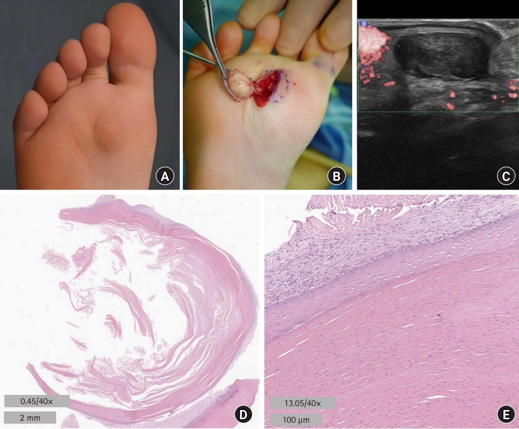
A 6-year-old female patient with a plantar epidermoid cyst (case 6). (A) Preoperative image. (B) Intraoperative image. (C) Preoperative ultrasonographic image showing avascular cystic lesion with heterogenous hypoechogenicity. (D, E) Microscopic images (H&E stain; scale bars, 2 mm in panel D and 100 μm in panel E).
There were three patients (cases 3, 5, and 8) with a history of multiple epidermoid cysts or benign soft-tissue tumor (nevus and lipoma), two with a history of cancer (thyroid cancer in case 1 and prostate cancer in case 2), and two with history of gastrointestinal polyposis (cases 2 and 5). Case 10 who demonstrated an early-onset epidermoid cyst in the lower extremity was diagnosed with Lowe syndrome.
All lesions were intraoperatively found in the subcutaneous layer. In nine of the 10 cases, the wounds were closed primarily after complete removal. However, in one case, malignant transformation to SCC arising from the cyst wall was incidentally found after the excision of the entire lesion with ulceration (case 5). After additional wide excision (including of the previous skin graft) with a margin of 2 cm, the resulting defect was reconstructed with a free thoracodorsal artery perforator (TDAP) flap (Fig. 2B).
In all cases, histopathology revealed epidermal cysts lined by stratified squamous epithelium and filled with cheese-like keratin in the laminated layers (Fig. 3D, 3E). Case 5 demonstrated multiple unilocular epidermoid cysts presenting as a single entity on the heel. In the largest cyst, the presence of a proliferating squamous epithelial portion of the cyst lining, with areas of cytonuclear atypia, indicated invasive well-differentiated SCC (Fig. 2C, 2D). No recurrence or complications were noted after surgery (mean follow-up, 8.7±4.4 years; range, 1–14 years), except in one case. In case 5, the epidermal cyst recurred twice (2 and 3 years after wide excision and reconstruction with the TDAP flap). Ruptured epidermal cysts on the flap were repeatedly excised, with no recurrence up to 6 years after the last surgery.
Discussion
Epidermal cysts are commonly caused by spontaneous or postinjury disruption of the pilosebaceous unit, leading to progressive cystic ectasia of the infundibular region of the hair follicle [1,6,16]. However, few studies have reported on the pathogenesis of epidermal cysts in non–hair-bearing regions, although Shimizu et al. [17] did report that most epidermal cysts were derived from traumatic sequestration of the keratinized squamous epithelium into the dermis. In our study, there was one case with a definite history of penetrating trauma to the palm (case 1). Case reports on epidermoid cysts that presumably resulted from chronic mechanical trauma such as shoe impingement or hammering have been suggested [8,18,19]. Conversely, several studies have reported HPV infection of the eccrine gland duct for possible etiology [9-12,20,21]. Although not performed in our study, tests for HPV infection, such as immunohistochemical staining and polymerase chain reaction may be considered in some cases.
Most epidermoid cysts are asymptomatic. Plantar epidermoid cysts can present with discomfort or pain during ambulation [6,22]. Firm swelling of the overlying hard skin of weight-bearing areas in the soles is usually protruding or palpable; however, one previously reported case did present with intractable heel pain caused by an epidermoid cyst, as revealed by magnetic resonance imaging [22]. Epidermoid cysts on the hand with increasing size cause pain resulting from pressure on the adjacent side. Once inflamed or ruptured, these cysts cause erythema, swelling, pain, and tenderness [1]. Incision and drainage can provide temporary relief; however, the lesions may eventually recur, as in the present cases 1, 2, and 5. Complete excision of the intact cyst wall via an elliptical incision with the inclusion of the central punctum is necessary to prevent a recurrence. In cases of active infection, surgical removal is delayed until the inflammation subsides after antibiotic therapy. The recurrence rate has been reported to be higher in the soles and palms than in other regions [12,16,23].
In general, differential diagnoses of epidermoid cysts in the hands and feet include warts, calluses, neuromas, ganglion cysts, fibromas, and lipomas [2,7]. The majority of epidermoid cysts are painless enlarging masses on the palmar aspect of the fingers. Previous reports have indicated the presence of a history of trauma in half of the patients, and more than half were manual laborers [23]. Areas that undergo repeated mechanical stress are exposed to chronic irritation and are at risk of recurrent minor traumas [19,24,25]. Case 1 also had a manual labor job. For plantar epidermoid cysts, the heel is the most common area, followed by the lateral border. In our study, however, the ball area was the most common region of the soles (three out of four patients).
Malignant transformation of epidermoid cysts is extremely rare (>1%) [14]. To date, only a few dozen cases of malignant transformation into SCC have been reported in the literature [13-15,26]. Although the precise mechanism remains unclear, the involvement of chronic inflammation due to remnant cystic components, especially in long-standing lesions, has been suggested to be involved in the general etiology of squamous cell carcinogenesis [15,26]. Similar to our case, most of these malignancies were detected incidentally on histopathological analysis after the excision of the whole lesion, which was presumed to be benign. The clinical features of suspected malignancy include rapid enlargement, ulceration of the overlying skin, prominent pain, or suspected infected cysts that fail to respond to medical therapy such as antibiotics [14,27,28].
Plantar epidermoid cysts in young children are rare [29]. Knight and Reincer [30] previously reported a greater incidence of plantar epidermoid cysts (10 out of 459 squamous cutaneous cysts) in children compared with that of adults (2.2% vs. 7.2%). In the pediatric population, the differential diagnoses for pediatric cases include pilomatricoma, dermoid cysts, and Gardner syndrome [1,32]. In this study, there were two pediatric cases with epidermoid cysts in the lower extremities (cases 6 and 10). In case 6, ultrasound imaging showing a well-circumscribed avascular cystic mass was useful in preoperative evaluation.
Most epidermoid cysts are sporadic; however, familial inheritance is possible. In particular, multiple and atypical localization of epidermoid cysts, such as limb involvement, may be indicative of familial syndromes. Case 10 diagnosed with rare, X-linked recessive Lowe syndrome presented with a relatively large epidermoid cyst in the lower extremity at the age of 4 years. This syndrome is mainly caused by a mutation of the OCRL1 gene. Recently, several studies have been reported on the associated skin lesions, such as multiple epidermoid cysts on the scalp [33]. On the other hand, case 6 was suspected of autosomal dominant Gardner syndrome with a mutation in the APC gene, which is characterized by multiple intestinal polyposis, osteomas, epidermoid cysts, and dental anomalies (e.g., multiple odontomas and supernumerary teeth) [4]. Early diagnosis based on these findings is important as malignant changes occur in colonic polyps that develop in the later decades of life.
Conclusion
In this study, we present a case series of epidermoid cysts found at unusual sites, such as the foot and hand. Differential diagnosis is important for these lesions through carefully taking a clinical history. The rapid growth of symptomatic epidermoid cysts with repeated recurrence may indicate malignant transformation. Complete removal of the mass, including the capsule, is important to prevent recurrence and histopathological confirmation. Once malignancy is confirmed, wide excision to achieve a safety margin should be performed, with the appropriate reconstruction of the resulting defect.
Notes
The authors have nothing to disclose.
Funding
None.