Surgical considerations for syndactyly of the hand in harlequin ichthyosis: a 5-year experience of sequential surgical treatment in a single patient
Article information

Abstract
Harlequin ichthyosis (HI) is a rare congenital disease that primarily affects the skin. Its complications include syndactyly, which can cause severe constriction. Surgical release is thought to be helpful, but the details remain unclear due to a lack of data. We share our 5-year experience of a patient with HI with successful outcomes from five syndactyly release operations involving division, local flap interposition, and full-thickness skin grafts. A visible decrease in the ulnar deviation of the metacarpophalangeal joints and a wider range of motion were noted. There were no complications, except for one case of donor site dehiscence that was resolved after minor revision. The surgical design must conform to the anatomical structure, as it can be easily mistaken due to masking of the skin. A suitable donor site is a flat, non-folding surface, such as the lateral thigh.
Introduction
Harlequin ichthyosis (HI) is a rare, autosomal recessive, severe genetic disorder that primarily affects the skin. It is characterized by hyperkeratosis and desquamation of the epidermidis, and patients are diagnosed at birth by their clinical appearance [1]. It is a skin disease with a wide range of complications. Neonates are born with excessive hyperkeratosis of the skin over nearly the entire body that forms deep, large, diamond-shaped plates [2,3]. This affects the shape of facial features and limits the movement of the arms and legs. The distal extremities are also affected, and joint contractures of the metacarpophalangeal (MCP) and interphalangeal (IP) joints, and syndactyly are among the features [4]. There is no cure for this condition, and supportive care; which includes moisturizing cream, antibiotics and retinoids are needed [5]. Due to the rarity of the disease, reports of satisfying surgical treatments of syndactyly are rare.
Four years ago, we reported a case of successful surgical treatment of syndactyly of a HI patient [6]. Satisfying results were noted and regular checkups were done. Sequential division surgeries were planned and done, and most recently the fifth surgery was performed. There were significant improvements both functionally and aesthetically with each surgery, and we would like to provide our experience for the appropriate surgical treatment and management of syndactyly of HI patients.
Case report
Ethics statement: The study was approved by the Institutional Review Board of Severance Hospital (No. 2022-0759-001) and performed in accordance with the principles of the Declaration of Helsinki. The patient’s legal guardian provided written informed consent for the publication and the use of the patient’s images.
A female patient diagnosed with HI at birth visited our outpatient department at the age of 6 years. Her skin showed severe erythroderma with generalized scaling. Her ears and nose were poorly developed, and ectropion was seen. Due to contraction of the skin, most of her joint movements were limited. She also showed syndactyly of both hands and feet. For the hand, all web spaces were involved, and skin contracture, ulnar deviation in proximal, middle, distal IP joints and MCP joints were noted.
Sequentially, the patient underwent five divisions, local flap interposition and full-thickness skin graft (Fig. 1, 2). The first surgery was performed on the right second interdigital space (IDS) when the patient was 6 years and 7 months old, and she showed functional improvement. She was able to manipulate a joystick and grasp paper. The next surgery was planned and done on the opposite left second IDS for the same effect. Then the third IDS was targeted to expand the range of motion and function. So, for the third and fourth surgery, the left and right third IDS was divided. The patient later showed recurred contraction on the left third IDS, and the latest fifth surgery was performed on this site for additional release. Specific details are shown in Table 1.
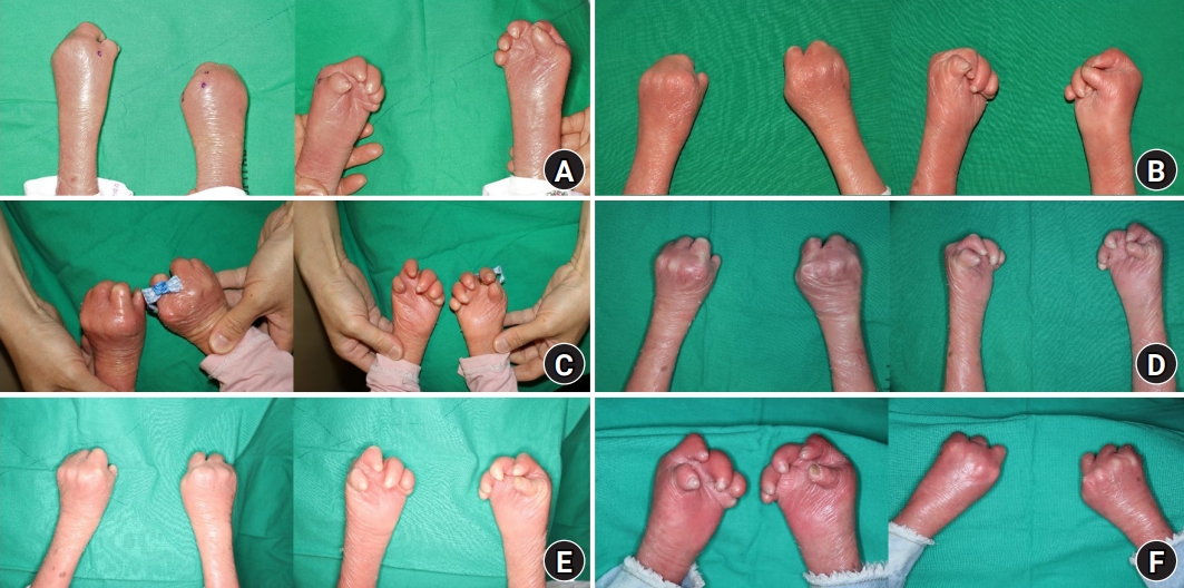
Sequential gross photographs. (A) Initial state at the age of 6 years and 7 months, showing incomplete syndactyly of all web spaces with skin contracture. (B) After the first operation (postoperative 6 months). (C) After the second operation (postoperative 5 months). (D) After the third operation (postoperative 5 months). (E) After the fourth operation (postoperative 10 months). (F) After the fifth operation (postoperative 1 month).

Sequential X-rays of the hands. (A) Initial state at the age of 6 years and 7 months, showing ulnar deviation in proximal, middle, and distal interphalangeal joints and metacarpophalangeal joints. (B) After the first operation (postoperative 1 year). (C) After the second operation (postoperative 4 months). (D) After the fourth operation (postoperative 9 months). For the ease of comparison, the right X-ray of Fig. 2A is reproduced from our previous report, Ahn et al. [6], according to the Creative Commons License.

Details of sequential operations
The same method was used for each surgery. Basically, the conventional syndactyly surgery method was followed; with the use of local flaps and full-thickness skin grafts [7]. A dorsal rectangular flap was designed all the way to the proximal PIP joint followed by a volar triangular flap, and they were drawn following the ulnar deviation of the MCP joints. It is important for the design to follow the anatomy as it can be misleading due to the masking caused by contracture and the skin (Fig. 3). The pneumatic tourniquet was inflated up to 200 mmHg. After division, contracture release around the incision was done. Then the dorsal rectangular flap and the volar triangular flaps were interposed to recreate the third web space, and leftover raw surface was covered with a full-thickness skin graft harvested from the lateral thigh, and primary repair for the donor site was done with #4-0 nylon. For the hand, suture was done with #4-0 and #5-0 vicryl and #5-0 nylon interruptedly (Fig. 4).

Photograph of intraoperative design of the fifth operation and preoperative X-ray of the left hand. (A) A surgical design that follows the anatomical shape and position of the metacarpus and proximal phalanxes. The yellow line shows a straight direction in which the alignment of the bones can be mistaken, and the red line shows the direction in which the bones are actually aligned. (B) X-ray view (inverted left and right).

Immediate postoperative photograph after the fifth operation. The left third web space is fully released with a full-thickness skin graft.
For the postoperative management, topical antibiotics was applied to the wound, moisturizer and oil were applied to the whole hand and compressive dressing with plain gauze and elastic bandage was maintained for about 1 week. The patient’s fingers were immobilized by tightly wrapping them with multiple layers of gauze, and no splinting was required. Only non-adherent dressing was used, and no direct taping was applied to the skin. After a week, open dressing with moisturizer and oil was done, and all stitches were removed 2 to 3 weeks after surgery. Flomoxef Na, a second-generation cephalosporin was given for 1 week. Postoperative stretching of the fingers was recommended 1 month after the surgery.
As for the donor site of the skin graft, the inguinal area was selected as in the conventional method. However, within 2 to 4 days, dehiscence occurred, and minor revision surgery was performed on the 12th day. Then on from the next surgery, the lateral thigh area was chosen as the skin graft donor site, and it healed well with no dehiscence or other complications (Fig. 5).
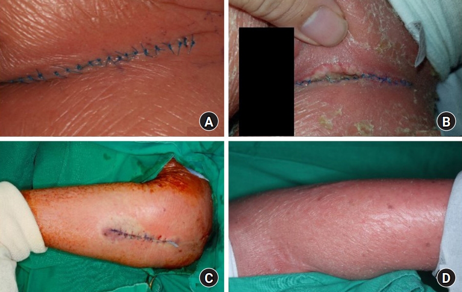
Healing of the donor site. Inguinal donor site after the first operation. (A) Postoperative 1 day. (B) Postoperative 10 days. Lateral thigh donor site after the second operation. (C) Immediate postoperative state. (D) Postoperative 3 years and 6 months, showing no signs of scarring.
After consecutive surgeries, visible decrease in the ulnar deviation of the MCP joints was noted on the X-ray (Fig. 1F, 2D). In particular, the patient was greatly satisfied with the easier operation of the joystick of her electric wheelchair, the ability to play with smaller toys and easily grip the pencil (Fig. 6).
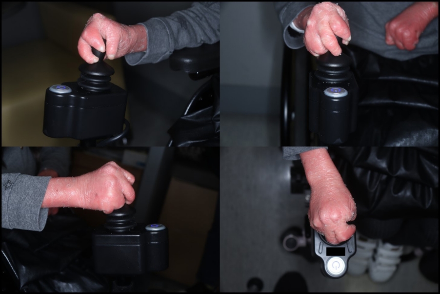
Photograph of the patient grasping the joystick of an electric wheelchair, at the age of 11 years, after 1 year and 7 months had passed since the fifth and last operation.
There were no other complications such as infection, dehiscence, delayed healing, hematoma, and seroma other than the donor site dehiscence after our first surgery that was mentioned above (Table 1) [6].
Discussion
HI is a rare genetic disorder with a short life expectancy in most cases. Thus, not much has been proven about this disease, and researches about syndactyly of HI patients are near to none [4]. However, recent advances in neonatal intensive care and coordinated multidisciplinary management have greatly improved survival rate and extended life expectancy [2,3]. With this development, syndactyly treatment for the patients’ improvement of quality of life should be considered.
Due to syndactyly, the patient cannot do a grasping motion and thus most activities are limited. However, after division surgery, movement of individual fingers and grasping became possible. Postoperative rehabilitation and stretching are thought to prevent worsening of contracture, but due to the patient’s young age and pain, compliance can easily fall low. So detailed explanations and encouragement to both the patient and their parents are needed.
When planning the surgery, the contracture and deviation of the hand is severe, so the surgical design must be done accordingly. This is because the actual bone structure may differ from what it looks like from the outside. The metacarpal bones and proximal phalanges can easily be mistaken because they are originally aligned in a straight line. However, in practice, it is necessary to recognize that each metacarpal bone and proximal phalanx are connected at an angle due to deviation, and design and surgery should be made accordingly.
The chosen skin graft donor for our first surgery was the left inguinal area. This is a common donor site for full-thickness skin graft and was thought to be a decent choice at that time [8]. However, as we explained in our previous report, dehiscence and delayed wound healing was noted [6]. Due to hyperkeratosis of the skin, elasticity was to a minimum resulting in no redundant skin. Also, because it is an area where the joint folds, when the patient spreads her leg, dehiscence of the wound worsened. Thus, the authors thought that skin harvest from a flat and non-folding area would be a better solution, and from the second surgery, the lateral thigh skin was used as the donor. This time, due to hyperkeratosis of the skin, scar healing with no complications and minimal scarring was noted. Therefore, although in our previous study we suggested that the skin graft size should be minimized, this is not necessarily the case. It is also important to note that the conventional skin graft donor site should not be used.
As for the timing of surgical intervention, it is not well known due to lack of studies. For otherwise normal patients, it is recommended that syndactyly release should be done from 3 to 24 months depending on the type and web space involved. Also, if the patient needs multiple surgeries, the timing has to be at least 3 months apart [9]. In our case, the patient was 6 years old when she was first examined in the outpatient department. At that time, contracture was already severe, and the authors thought that there would have been better results if the surgery had been performed earlier. In one study, it is said that for HI patients, it would be good to relieve the contracture with surgeries from the beginning at birth [10]. We give a similar opinion, that surgery should be done from 3 to 24 months, and regarding the surgery interval, 3 months is enough time for recovery.
Surgical intervention will always increase the possibility of infection, and in HI, such an infection is dangerous and can easily lead to a systemic infection, which is the leading cause of death in HI [5]. So thorough use of intravenous and topical antibiotics is thought to be important. However, since there is no consensus in the literature, prophylactic antibiotics used in general patients were administrated. A second-generation cephalosporin was given for a week, and no infection occurred for all surgeries.
There are several limitations of this study. First, due to the rarity of the disease, we had no choice but to look at only one case, and direct comparison with other cases was not possible. Second, due to its retrospective nature, the hands were monitored only via X-rays and observational windows, which could be subjective and questionable. So, in the future, we shall check on objective results such as the DASH (Disabilities of Arm, Shoulder and Hand) score. Third, with a 5-year follow-up period, it is difficult to know the future results and prognosis, and longer follow-ups are needed.
In conclusion, for patients with HI, proper syndactyly release with full-thickness skin graft is important and can be done safely. Surgical design should follow the anatomical shape and position of the bones and soft tissue. A flat non-folding surface, such as the lateral thigh should be chosen as the skin graft donor site. Multiple surgeries with sufficient time intervals should also be considered for further improvements.
Notes
The authors have nothing to disclose.
Funding
None.