Arch Hand Microsurg Search

CLOSE


Arch Hand Microsurg > Volume 25(1); 2020 > Article
척골충돌증후군의 수술 중 관절경 소견

Abstract

Purpose

The purpose of this study was to compare the preoperative magnetic resonance imaging (MRI) findings and intraoperative arthroscopic findings in ulnar impaction syndrome, and to assess the necessity of the arthroscopic evaluation in ulnar shortening osteotomy.

Methods

We retrospectively reviewed 46 patients of idiopathic ulnar impaction syndrome, treated by ulnar shortening osteotomy and arthroscopic evaluation, from February 2012 to June 2018. In preoperative MRI and intraoperative arthroscopy, lunate, triquetrum, triangular fibrocartilage complex (TFCC) and lunotriquetral ligament were evaluated.

Results

The chance to see the findings of chondromalacia of the lunate and triquetral cartilage in arthroscopy (58% and 2%) was less than the findings of abnormal signal intensity of the lunate and triquetrum in MRI (65% and 22%). The incidence of TFCC lesion in arthroscopy (89%) was almost equal with that of abnormal signal intensity of TFCC in MRI findings (87%). But there were 24 cases (52%) where the prediction of triangular fibrocartilage lesions in MRI was incorrect. In 19 cases (41%), arthroscopic findings were not appropriate with the disease stage of Palmer’s original classification.

Conclusion

Although some recent reports say that arthroscopic surgery is not necessary in the treatment of ulnar impaction syndrome, we could confirm the value of arthroscopic evaluation for the identification of ulnocarpal pathologies. We would like to recommend the arthroscopic evaluation of ulnocarpal joint at the same time of the ulnar shortening procedure.

서론

척골충돌증후군은 척측 수근관절 동통을 유발하는 흔한 원인 중 하나로 단순방사선검사에서 척골의 양성 변위를 보이며, 척측 수근관절의 반복적 부하로 인해 삼각섬유연골복합체와 척측 수근골의 퇴행성 변화를 유발한다[1]. 척골충돌증후군의 수술적 치료방법으로는 척골 단축술이 보편적인 치료방법으로 널리 시행되고 있으며, 관절경 시술의 발달로 척골 단축술과 수근관절 관절경을 같이 시행하는 방법도 시행되고 있다[2-6]. 척골충돌증후군과 삼각섬유연골복합체의 손상이 동반되는 경우, 손상된 삼각섬유연골복합체의 불안정한 파열판은 척골 두와 척측 수근골 사이에서 기계적인 자극을 유발할 수 있어 변연절제술을 시행하는 것이 유용할 수 있다[5,7]. 하지만 척골 단축술과 관절경적 변연절제술을 동시에 시행하는 경우, 관절경 시술의 효용성에 대해서는 아직 논란의 여지가 있다.
척골충돌증후군에서 삼각섬유연골복합체와 척측 수근골의 손상 등의 관절내 병변의 진단으로 자기공명영상과 관절경 등이 보고되었으나, 삼각섬유연골복합체의 손상, 척측 수근골의 연골연화증, 월상삼각골간 인대의 손상의 정확한 진단에 자기공명영상은 한계가 있는 것으로 지적되고 있다[6,8-10]. 따라서 관절경을 시행하여 척측 수근관절 내부의 병변을 확인하고 수술방법을 결정하는 것이 유용할 것으로 생각하였다.
저자들은 척골충돌증후군으로 진단된 환자들에 대해 수근관절의 관절경을 시행하였으며, 삼각섬유연골복합체의 손상 및 척측 수근골의 연골연화증 등의 관절내 병변을 확인하고, 관절경적 변연절제술과 척골 단축술을 동시에 시행하였다. 수술 전 시행한 자기공명영상검사 소견과 수술 중 확인된 관절경 소견을 후향적으로 분석하여, 자기공명영상검사 소견과 관절경 소견을 비교하고, 척골충돌증후군의 관절경 소견을 분석하여, 척골 단축술과 동시에 시행하는 관절경 시술의 효용성을 알아보고자 하였다.

대상 및 방법

2012년 2월부터 2018년 6월까지 척골충돌증후군으로 척골 단축술과 수근관절 관절경적 변연절제술을 받은 환자 중, 수술 전 자기공명영상을 촬영하였고, 1년 이상 추시 관찰이 가능하였던 46예를 대상으로 하였다. 평균 추시 기간은 20.5개월(범위, 12-28개월)이였고 남자가 21예, 여자가 25예였으며, 좌측이 20예, 우측이 26예이었다. 평균 연령은 49.2세(범위, 21-75세)였다. 증상 발생일로부터 수술을 받기까지의 기간은 평균 21.5개월(범위, 5-60개월)이었다.
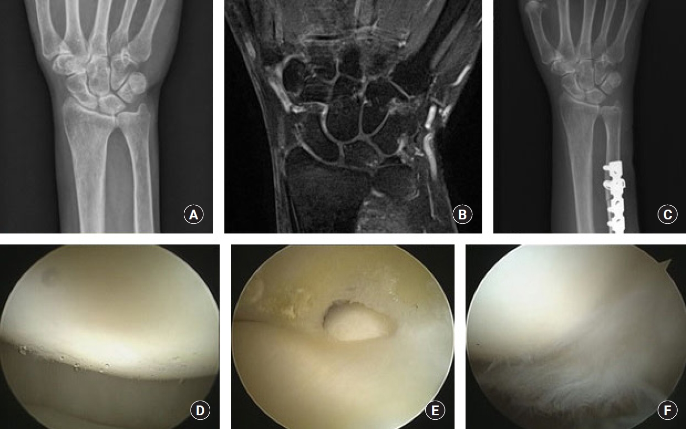
척측 수근관절 동통을 호소하는 환자 중 진찰 소견상 척골 두와 삼각골, 월상골 사이 관절면에 집중된 압통이 있으며, 척수근부하검사(ulnocarpal stress test) 및 척수근연마검사(ulnocarpal grind test)상 양성 소견을 보인 환자들을 척골충돌증후군으로 진단하였으며[7] 본원에 내원 후 최소 3개월 이상 수근관절 스트레칭 물리치료 및 약물로 보존적 치료를 시행하였다. 3개월 이상의 보존적 치료로 증상 완화에 실패한 환자를 대상으로 3.0T 자기공명영상촬영을 하였다. 이학적 검사상 양성 소견이 지속되며, 단순방사선촬영상 양성 척골 변위나 월상골의 미란이나 경화 소견을 보이거나 자기공명영상촬영상 월상골, 삼각골의 골수 내 신호강도의 변화나 삼각섬유연골의 퇴행성 병변을 보이는 환자를 대상으로 요척수근관절 관절경 및 척골 단축술을 시행하였다. 이학적 검사 및 자기공명영상검사상 척골충돌증후군으로 의심되나 척골 양성 변이를 보이지 않는 환자들은 관절경적 변연절제술만 시행하였고 보존적 치료 시행 후 통증 호전은 없으나 기능적으로 우수한 환자들은 대부분 보존적 치료를 유지하였고 대상에서 제외하였다. 수술 전후 단순 후전방 방사선사진에서 Kreder 등[11]의 계측방법을 사용하여 척골의 양성 변위를 측정하였고, 수술 전 자기공명영상에서 변형된 Outerbridge 분류로 월상골과 삼각골의 비정상신호 음영 여부 및 연골결손을 분류하고 삼각섬유연골복합체의 손상 유무, 월상삼각골간 인대의 손상 유무를 관찰하였다.
수술시에는 전신 마취 하 지혈대를 착용하고, 요척수근관절에 대한 관절경 시술을 먼저 시행하였다. 3-4 관절경 입구를 통해 관절경을 이용하여 변형된 Outerbridge 분류로 월상골과 삼각골의 연골연화증을 분류하고 삼각섬유연골복합체와 월상삼각골간 인대의 손상, 척골 경상돌기 전 함몰부의 활액막염을 확인하였다. 삼각섬유연골복합체의 손상은 Palmer [12]의 분류에 따라 분류하였다. 6R 관절경 입구를 통해 전동 소파기와 고주파 열치료기(ArthroCare, Austin, TX, USA)를 이용하여 삼각섬유연골복합체와 월상삼각골간 인대의 손상, 척골 경상돌기 전 함몰부의 증식된 활액막에 대하여 변연절제술을 시행하였다. 관절경 시술 후 관절경 입구의 피부를 봉합한 후, 척골의 척측연을 따라 6-7 cm 정도의 피부 절개를 가하고, 척 수근신근과 척 수근굴근의 사이로 척골에 도달하였다. 척 수근관절에서 근위 4-5 cm 되는 부위에 수술 전 방사선사진에서 측정한 양성 척골 변위의 정도를 고려하여 수술 전 계산된 절제량을 표시하였다. 전동톱(oscillating saw)으로 사선형 절골술을 시행하고 단축술 후 척골의 변위가 0-1 mm가 되도록 사선형으로 절골하여 단축시켰다. 절골 부위는 5 혹은 6 hole의 3.5 mm 잠금 압박 금속판을 척골의 배부에 고정하였다. 수술 후 설탕집게 부목으로 약 4주간 절대 고정하였고, 이후에는 제거 가능한 보조기로 간헐적 운동을 허용하였다.

결과

수술 전 시행한 자기공명영상검사에서 31예(67%)에서 월상골의 연골연화 소견을 보였고, Grade I이 7예, Grade II가 21예, Grade III이 3예였다. 10예(22%)에서 삼각골의 연골연화 소견을 보였고, Grade I이 8예, Grade II가 2예였다. 42예(91%)에서 삼각섬유연골복합체의 비정상신호 음영 소견을 보였으며, 이중 20예에서 마모 등의 퇴행성 변화가 의심되었고, 22예에서 파열이 의심되었다. 월상삼각골간 인대의 손상은 1예(2%)에서 의심되었다.
요척수근관절경에서 28예(60%)에서 월상골의 연골연화 소견을 보였고, Grade II가 24예, Grade III이 1예, Grade IV가 3예였다(Fig. 1). 삼각골의 연골연화 소견은 1예(2%)에서 Grade II로 관찰되었다. 43예(94%)에서 삼각섬유연골의 손상이 관찰되었으며, 이중 11예에서 퇴행성 마모가 관찰되었고, 32예에서 파열이 관찰되었다. 삼각섬유연골의 파열이 관찰된 32예 중 25예에서 중앙부 파열, 6예에서 요측 파열, 1예에서 척측 파열이 관찰되었다. 11예(24%)에서 월상삼각골간 인대의 손상이 관찰되었으며, 이중 7예에서 퇴행성 마모, 4예에서 피판 파열 소견을 보였다(Table 1).
자기공명영상검사와 관절경 소견을 비교하면, 월상골은 11예에서 자기공명영상검사상 6예의 Grade I, 3예의 Grade II, 1예의 Grade III의 연골연화 소견을 보였으나 관절경상 관절 연골의 정상소견을 보였고, 7예에서 자기공명영상검사상 정상이었으나 관절경에서 Grade II의 연골연화 소견을 보여 차이를 보였다. 삼각섬유연골복합체는 24예가 자기공명영상검사 소견과 관절경 검사 소견의 차이를 보였다. 자기공명영상검사에서 파열이 의심되었던 8예에서 관절경에서는 5예의 마모 소견과 3예의 정상 소견을 보였고, 자기공명영상검사에서 마모가 의심되었던 14예에서 관절경에서는 13예의 파열 소견과 1예의 정상 소견을 보였다(Fig. 2). 2예에서는 자기공명영상검사에서 중앙부 파열이 의심되었으나, 관절경에서는 요측 파열 소견을 보였다. 월상삼각골간 인대는 관절경에서 손상을 보였던 11예는 자기공명영상검사에서는 정상 소견을 보였다.
관절경 검사 소견에 의한 Palmer [12] 분류는 2A형이 2예, 2B형이 3예, 2C형이 15예, 2D형이 6예, 2E형이 1예였다. 19예에서 Palmer [12]의 분류와 일치하지 않는 소견을 보였는데, 17예에서 월상골의 연골연화 소견 없이 삼각섬유연골의 마모 및 파열 소견을 보였고(Fig. 3), 2예에서 삼각섬유연골의 파열 소견 없이 월상삼각골간 인대의 마모 및 파열 소견을 보였다(Table 2).
척골 변위는 술 전 3.8 mm (범위, 1.2-6 mm)에서 술 후 0.2 mm (범위, -1.2-1.6 mm)로 교정되었으며, 불유합이나 고정 실패를 보인 예는 없었다. 평균 13.2개월(범위, 11-15개월)에 전 예에서 기구제거 수술을 시행하였으며, 수술과 관련된 합병증이 발생한 환자는 없었다.

고찰

척골충돌증후군은 척측 수근관절의 월상골, 삼각골, 척골 두, 삼각섬유연골복합체, 월상삼각골간 인대에 대한 반복적 부하로 인해 발생하는 퇴행성 질환으로, 월상골의 연골연화증과 삼각섬유연골복합체의 마모에서부터 퇴행성 변화가 시작되는 것으로 알려져 있다[1]. 퇴행성 변화가 진행되면 삼각골 및 척골 두의 연골연화증이 발생하고, 삼각섬유연골복합체는 천공이 되며, 더 진행 시 월상삼각골간 인대의 파열까지 발생한다[13,14].
척측 수근관절 동통을 호소하는 환자에서 진찰 소견상 척골충돌증후군이 의심되면 자기공명영상검사를 시행할 수 있다. 자기공명영상검사에서는 월상골, 삼각골, 척골 두의 신호강도 변화 및 연골결손을 확인하고, 삼각섬유연골복합체 및 월상삼각골간 인대손상을 확인 또는 의심할 수 있다. 그러나 자기공명영상검사에서 확인된 월상골의 신호강도 변화로 연골연화증의 정도를 감지하는 것은 힘들고[8], 삼각섬유연골복합체의 마모 및 파열, 월상삼각골간 인대의 병변을 감지하는 것에도 한계가 있다[13,15]. Hobby 등[16]은 자기공명영상검사에서 삼각섬유연골복합체 손상의 진단의 정확도를 88%로 보고하였고 월상삼각골간 인대손상의 진단의 정확도는 82%로 보고하였다. Magee [17]는 관절경 검사에서 진단된 22예의 삼각섬유연골복합체 손상 환자 중 19예가 자기공명영상검사에서 확인되었고(86%), 관절경 검사에서 진단된 11예의 월상삼각골간 인대손상 환자 중 9예가 자기공명영상검사에서 확인되었다고(81%) 보고하였다. 척골충돌증후군에서 시행한 자기공명영상검사상 삼각섬유연골복합체의 손상이 관절경 소견과 일치하지 않는 결과가 여러 저자들에 의해 보고되었다[3,6]. 본 연구에서도 자기공명영상검사 소견과 관절경 소견이 일치하지 않았던 환자들을 관찰할 수 있었다. 특히 자기공명영상검사상 월상골과 삼각골에서 관찰되는 골수의 신호강도 변화와 관절경에서 관찰되는 연골 소견과 차이를 보이고 있어 골수의 신호강도 변화는 연골연화증의 초기 소견으로 생각되며 자기공명영상에서 관찰되는 신호강도의 변화로 연골연화증의 정도를 평가하는 것은 한계가 있을 것으로 생각된다. 삼각섬유연골복합체도 24예(52%)에서 일치하지 않았으며 특히 마모 소견을 보였던 14예 중 13예가 파열 소견을 보여 변연절제술을 시행하였다. 월상삼각골간 인대손상이 있었던 11예는 자기공명영상에서 진단이 되지 않았다. 척골충돌증후군과 동반된 관절내 병변의 진단 시 자기공명영상검사의 정확도가 비교적 높은 편이지만 관절경 소견과 차이를 보이고 있어, 정확한 진단 및 치료의 결정을 위해서는 관절경 검사의 역할이 더 높게 평가되어야 한다고 생각된다.
최근의 연구결과에서 단순방사선검사와 자기공명영상검사를 기반으로 한 Palmer [12]의 삼각섬유연골복합체 손상 분류에 의문이 제기되고 있다. Koh 등[18]은 50예의 척골충돌증후군 환자에서 관절경 소견상 21예(42%)에서 월상골의 연골연화증 없이 삼각섬유연골의 손상을 보이거나 삼각섬유연골의 손상 없이 월상삼각골간 인대의 손상을 보여 Palmer [12]의 분류와 일치하지 않는 소견을 보고하였다. 본 연구에서도 46예 중 19예(41%)의 관절경 소견이 Palmer [12]의 분류와 일치하지 않는 소견으로 비슷한 결과를 보였다. 하지만 환자군 선정 시 외상력을 제외기준으로 설정하지 않았고, 3예의 요측 파열과 1예의 척측 파열도 포함되어 있어 간과된 외상성 삼각섬유연골과 월상삼각골간 인대의 손상 가능성도 고려해야할 것으로 생각된다. 관절경 술식의 발달로 삼각섬유연골복합체 및 요척수근관절 내 병변에 대한 더 정확한 검사가 가능해지면서 척골충돌증후군에서 병변의 진행에 대한 분류와 외상성 병변의 감별에 대한 추가적 연구가 필요할 것으로 생각된다.
관절경 술식의 발달로 척골 단축술에서 관절경 시술을 동시에 시행하는 방법도 많이 사용되고 있다[3,4,7,18-20]. 관절경 검사 시 삼각섬유연골복합체의 손상 및 수근골의 연골연화증을 더 정확하게 관찰할 수 있고[3], 요척수근관절 병변의 진단 및 변연절제술 등의 수술적 처치를 동시에 시행할 수 있는 장점이 있다[4,6]. 하지만 척골 단축술과 관절경 시술을 동시에 시행하였을 때 관절경 시술이 치료결과에 미치는 영향에 대해서는 아직 논란의 여지가 있다. Roh 등[20]은 관절경적 변연절제술을 동시에 시행할 경우 단기 추시 시 통계적으로 유의한 통증의 감소가 있었으나 장기 추시 시에는 통증과 임상적 결과의 유의한 차이가 없음을 보고하였고, Kim 등[5]과 Park 등[19]은 관절경적 변연절제술의 병행 여부는 척골 단축술의 치료결과에 영향을 미치지 않으므로 모든 환자에게 관절경 시술을 병행할 필요는 없다고 하였다. 하지만 위 연구들은 척골충돌증후군에서 척골 단축술과 함께 시행한 관절경적 변연절제술의 임상적 치료결과만을 비교한 것으로 관절경 검사에서 관찰되는 개별 병변에 대한 치료효과도 척골충돌증후군 치료 시 고려해야할 점으로 생각된다. 삼각섬유연골복합체의 파열에 대해 Tatebe 등[21]은 척골 단축술 후 낮은 DASH 점수와 연관이 있다고 하였고, Low 등[22]과 Hulsizer 등[23]은 척골충돌증후군에서 삼각섬유연골복합체의 변연절제술만으로 증상 호전을 보고하였다. 하지만 척골충돌증후군에서 삼각섬유연골복합체 파열의 관절경적 변연절제술을 단독으로 시행하여 좋지 않은 결과도 보고되고 있어[5] 아직 논란의 여지가 있다. Minami 등[10]은 척골 중립 및 음성 변위를 보이는 삼각섬유연골복합체 손상에서는 변연절제술만으로 우수한 결과를 보였지만, 척골 양성 변위를 보이는 삼각섬유연골복합체 손상에서는 불량한 결과를 보임을 보고하였다. 따라서 척골 양성 변위를 보이는 척골충돌증후군에서 삼각섬유연골복합체의 변연절제술만으로 치료하는 것은 한계가 있으며 척골 단축술과 동시에 시행 시 치료결과에 미치는 영향에 대해서 추가적인 연구가 필요할 것으로 생각된다. Koh 등[18]은 삼각섬유연골복합체의 중심성 파열, 월상골 연골의 불안정한 피판 파열, 월상삼각골간 인대의 피판 파열과 같은 관절 내 병변은 관절경적 변연절제술이 도움이 된다고 주장하였다. 월상삼각골간 인대의 손상은 부분 및 피판 파열 시 변연절제술의 우수한 치료결과가 여러 저자들에 의해 보고되고 있어 손상이 관찰되면 변연절제술을 시행하는 것이 치료에 도움이 될 것으로 생각된다[16,24-26]. 척골 단축술의 우수한 결과들로[3-6,13,15] 척골 단축술이 척골충돌증후군의 주된 치료방법으로 널리 사용되고 있으나, 관절경에서 관찰되는 관절 내 병변에 대한 변연절제술 등의 술식도 치료에 도움을 줄 것으로 생각된다.
본 연구의 한계로는 증례수가 46예로 비교적 적었고, 추시 기간이 평균 20.5개월로 짧아 장기적으로 발생할 수 있는 관절염의 발생 유무 등에 대하여 알기 힘든 점 등이 있었다. 본 연구에서는 관절경 시행 시 요척수근관절만 관찰하였다. 추가적으로 요척관절, 중수근관절 관절경을 시행 시 더 정확한 진단이 가능하며 추가적인 처치가 필요할 수 있을 것으로 생각된다. 또한 관절경 술식에서 변연절제술 이외의 치료가 필요한 관절내 병변은 보이지 않았고, 척골충돌증후군에서 발견되는 관절내 병변에 대한 변연절제술이 척골충돌증후군의 치료결과에 미치는 영향을 평가하기에는 부족하여, 이를 보완하기 위해 더 많은 수의 증례를 대상으로 장기 추시 연구가 필요할 것으로 생각된다.

결론

척골충돌증후군 치료에서 수술 전 평가로 자기공명영상검사가 발전되었지만, 관절경 술식은 척수근 관절의 병변을 직접 관찰하여 보다 정확한 진단이 가능한 장점이 있다. 척골 단축술은 척골충돌증후군에서 우수한 치료결과를 얻을 수 있는 유용한 치료법으로 생각된다. 단, 관절경적 변연절제술의 병행 여부가 척골충돌증후군의 치료결과에 미치는 영향에 대해서는 추가적인 연구가 필요하다고 생각된다.

Conflicts of interest

The authors have nothing to disclose.

Fig. 1.
A 60-year-old female patient complained of right wrist ulnar side pain. (A) Preoperative radiograph shows positive ulnar variance (3.72 mm). (B, C) Magnetic resonance imaging shows subchondral cysts in the lunate and triquetrum and tear of the triangular fibrocartilage (TFC). (D) Postoperative radiograph shows corrected ulnar variance (0.00 mm). (E) Arthroscopic view from the 3–4 portal showing severe defect of the articular cartilage of the lunate. (F) Arthroscopic image showing a perforation of the TFC.
ahm-19-0067f1.jpg
Fig. 2.
A 76-year-old female patient complained of left wrist ulnar side pain. (A) Pre-operative radiograph shows positive ulnar variance (5.43 mm). (B) Magnetic resonance imaging (MRI) shows degeneration of the TFC. (C) Post-operative radiograph shows corrected ulnar variance (-0.86 mm). (D) Arthroscopic view from the 3–4 portal showing degenerative wear of the articular cartilage of the lunate. (E) Arthroscopic image showing a perforation of the triangular fibrocartilage (TFC). (F) Arthroscopic image showing degenerative wear of the lunotriquetral interosseous ligament. MRI shows degeneration of the TFC, but arthroscopic finding shows a perforation of the TFC.
ahm-19-0067f2.jpg
Fig. 3.
A 77-year-old female patient complained of right wrist ulnar side pain. (A) Pre-operative radiograph shows positive ulnar variance (3.19 mm). (B) Magnetic resonance imaging shows multifocal subchondral bone edema of the lunate and degeneration of the triangular fibrocartilage complex (TFCC). (C) Post-operative radiograph shows corrected ulnar variance (-1.22 mm). (D) Arthroscopic view from the 3–4 portal showing intact articular cartilage of the lunate. (E) Arthroscopic image showing a degenerative central tear of the triangular fibrocartilage. (F) Arthroscopic image showing a flap tear of the lunotriquetral interosseous ligament. Arthroscopic findings were not appropriate with Palmer’s original description of TFCC.
ahm-19-0067f3.jpg
Table 1.
Preoperative MRI findings and Intraoperative arthroscopic findings
MRI findings Arthroscopic findings
Lunate chondromalacia 31 (67) 28 (60)
 Grade I 7 -
 Grade II 21 24
 Grade III 3 1
 Grade IV - 3
Triquetrum chondromalacia 10 (22) 1 (2)
 Grade I 8 -
 Grade II 2 1
TFCC lesion 42 (91) 43 (94)
 Wear 20 11
 Perforation 22 32
  Central 25
  Radial 6
  Ulnar 1
LT ligament lesion (tear) 1 (2) 11 (24)
 Wear 7
 Flap tear 4

Values are presented as number (%).

MRI, magnetic resonance imaging; TFCC, triangular fibrocartilage complex; LT, lunotriquetral.

Table 2.
Patient demographics, MRI and arthroscopic findings
Case Sex/Age (yr) MRI Arthroscopy Palmer’s classification (arthroscopy)
Lunate grade Triquetrum grade TFCC LTIL Lunate grade Triquetrum grade TFCC LTIL
1 F/71 II Normal Central tear Intact Normal Normal Radial tear Tear with flap
2 M/40 II Normal Central tear Intact II (fibrillation) Normal Central tear with flap Intact 2C
3 M/65 II I Central tear Intact II (fibrillation) Normal Wear (thinned) Intact 2B
4 F/34 Normal Normal Central tear Intact II (fibrillation) Normal Large perforation Intact 2C
5 M/50 Normal Normal Central tear Intact II (fibrillation) Normal Central tear Wear 2D
6 M/36 III I Central tear Intact II (fibrillation) Normal Wear (thinned) Wear 2D
7 F/70 II Normal Degeneration Intact III (cartilage flap) Normal Radial tear Intact 2C
8 F/38 II Normal Degeneration Intact II (fibrillation) Normal Central tear Intact 2C
9 M/59 Normal Normal Central tear Intact Normal Normal Wear (thinned) Intact
10 M/21 Normal Normal Central tear Intact II (fibrillation) Normal Wear (thinned) Intact 2B
11 M/25 I Normal Central tear Intact Normal Normal Central tear Intact
12 F/55 III I Degeneration Intact IV (chondral defect) Normal Central tear Intact 2C
13 M/44 II Normal Degeneration Intact II (fibrillation) Normal Central tear Intact 2C
14 M/41 II Normal Degeneration Intact II (fibrillation) Normal Central tear Intact 2C
15 F/64 II I Central tear Intact II (fibrillation) Normal Intact Intact 2C
16 F/33 Normal I Central tear Intact Normal Normal Central tear Intact
17 M/75 II I Central tear Intact Normal Normal Central tear Intact
18 M/72 I II Central tear Intact Normal Normal Central tear Intact
19 F/54 Normal Normal Intact Intact Normal Normal Large perforation Wear
20 F/48 III Normal Central tear Intact Normal Normal Radial tear Intact
21 F/53 I Normal Central tear Intact Normal Normal Central tear Intact
22 F/41 Normal Normal Partial tear Intact Normal Normal Small tear Intact
23 M/28 Normal Normal Degeneration Intact II (fibrillation) Normal Radial tear Intact 2C
24 M/55 II Normal Degeneration Intact IV (chondral defect) Normal Central tear Intact 2C
25 F/63 II Normal Degeneration Intact II (fibrillation) Normal Wear (thinned) Wear
26 F/58 I Normal Degeneration Intact Normal Normal Small tear Intact
27 F/52 Normal Normal Degeneration Intact Normal Normal Radial tear Intact
28 M/59 II II Central tear Intact II (fibrillation) II (fibrillation) Wear (thinned) Wear 2D
29 F/56 II Normal Central tear Tear II (fibrillation) Normal Central tear Intact 2C
30 F/54 II Normal Degeneration Intact II (fibrillation) Normal Wear (thinned) Tear with flap
31 M/60 II I Degeneration Intact II (fibrillation) Normal Wear (thinned) Tear with flap 2D
32 M/47 II Normal Central tear Intact II (fibrillation) Normal Central tear Wear 2D
33 F/57 Normal Normal Intact Intact II (fibrillation) Normal Central tear Wear 2D
34 M/47 II I Degeneration Intact II (fibrillation) Normal Central tear Partial tear with flap 2E
35 M/49 II Normal Intact Intact IV (chondral defect) Normal Central tear Intact 2C
36 F/56 Normal Normal Central tear Intact Normal Normal Central tear Intact
37 F/49 II Normal Intact Intact II (fibrillation) Normal Intact Intact 2A
38 F/33 II Normal Degeneration Intact II (fibrillation) Normal Intact Intact 2A
39 F/53 Normal Normal Degeneration Intact II (fibrillation) Normal Wear (thinned) Intact 2B
40 M/44 Normal Normal Central tear Intact II (fibrillation) Normal Central tear Intact 2C
41 F/30 Normal Normal Degeneration Intact Normal Normal Central tear Intact 2C
42 F/49 II Normal Degeneration Intact Normal Normal Ulnar tear Intact
43 F/46 I Normal Central tear Intact Normal Normal Central tear Intact
44 M/31 I Normal Degeneration Intact Normal Normal Degeneration Intact
45 F/49 Normal Normal Degeneration Intact Normal Normal Severe degeneration Intact
46 M/49 I Normal Degeneration Intact II (fibrillation) Normal Radial tear Intact 2C

MRI, magnetic resonance imaging; TFCC, triangular fibrocartilage complex; LTIL, lunotriquetral interosseous ligament.

References

1. Friedman SL, Palmer AK. The ulnar impaction syndrome. Hand Clin. 1991;7:295-310.
crossref pmid
2. Bernstein MA, Nagle DJ, Martinez A, Stogin JM Jr, Wiedrich TA. A comparison of combined arthroscopic triangular fibrocartilage complex debridement and arthroscopic wafer distal ulna resection versus arthroscopic triangular fibrocartilage complex debridement and ulnar shortening osteotomy for ulnocarpal abutment syndrome. Arthroscopy. 2004;20:392-401.
crossref pmid
3. Cheon SJ, Kang JH, Cho JY, Lim JM. Treatment of ulnar impaction syndrome using arthroscopy and ulnar shortening osteotomy. J Korean Orthop Assoc. 2007;42:310-7.
crossref
4. Park JW, Kim SK, Park JH, Wang JH, Lee SJ, Jeon WJ. Results of Ulnar Shortening for Ulnar Impaction Syndrome in the Wrist. J Korean Soc Surg Hand. 2006;11:14-9.

5. Kim BS, Song HS. A comparison of ulnar shortening osteotomy alone versus combined arthroscopic triangular fibrocartilage complex debridement and ulnar shortening osteotomy for ulnar impaction syndrome. Clin Orthop Surg. 2011;3:184-90.
crossref pmid pmc
6. Baek GH, Chung MS, Lee YH, Jeong GI, Lee CH. Arthroscopy of the wrist and ulnar shortening osteotomy for the treatment of the ulnar impaction syndrome. J Korean Orthop Assoc. 2001;36:207-14.
crossref
7. Sachar K. Ulnar-sided wrist pain: evaluation and treatment of triangular fibrocartilage complex tears, ulnocarpal impaction syndrome, and lunotriquetral ligament tears. J Hand Surg Am. 2008;33:1669-79.
crossref pmid
8. Imaeda T, Nakamura R, Shionoya K, Makino N. Ulnar impaction syndrome: MR imaging findings. Radiology. 1996;201:495-500.
crossref pmid
9. Escobedo EM, Bergman AG, Hunter JC. MR imaging of ulnar impaction. Skeletal Radiol. 1995;24:85-90.
crossref pmid pdf
10. Minami A, Ishikawa J, Suenaga N, Kasashima T. Clinical results of treatment of triangular fibrocartilage complex tears by arthroscopic debridement. J Hand Surg Am. 1996;21:406-11.
crossref pmid
11. Kreder HJ, Hanel DP, McKee M, Jupiter J, McGillivary G, Swiontkowski MF. X-ray film measurements for healed distal radius fractures. J Hand Surg Am. 1996;21:31-9.
crossref pmid
12. Palmer AK. Triangular fibrocartilage complex lesions: a classification. J Hand Surg Am. 1989;14:594-606.
crossref pmid
13. Chun S, Palmer AK. The ulnar impaction syndrome: follow-up of ulnar shortening osteotomy. J Hand Surg Am. 1993;18:46-53.
crossref pmid
14. Cerezal L, del Pinal F, Abascal F, Garcia-Valtuille R, Pereda T, Canga A. Imaging findings in ulnar-sided wrist impaction syndromes. Radiographics. 2002;22:105-21.
crossref pmid
15. Loh YC, Van Den Abbeele K, Stanley JK, Trail IA. The results of ulnar shortening for ulnar impaction syndrome. J Hand Surg Br. 1999;24:316-20.
crossref pmid
16. Hobby JL, Tom BD, Bearcroft PW, Dixon AK. Magnetic resonance imaging of the wrist: diagnostic performance statistics. Clin Radiol. 2001;56:50-7.
crossref pmid
17. Magee T. Comparison of 3-T MRI and arthroscopy of intrinsic wrist ligament and TFCC tears. AJR Am J Roentgenol. 2009;192:80-5.
crossref pmid
18. Koh KH, Lee HL, Chang YS, Park MJ. Arthroscopy during ulnar shortening for idiopathic ulnar impaction syndrome. Orthopedics. 2013;36:e1495-500.
crossref pmid
19. Park JC, Sur YJ, Rhee SK, Song SW, Lee SM, Han SH. Ulnar shortening osteotomy for the treatment of ulnar impaction syndrome. J Korean Soc Surg Hand. 2009;14:172-8.

20. Roh YH, Song JH, Gong HS, Baek GH. Comparison of clinical outcomes after ulnar shortening osteotomy for ulnar impaction syndrome with or without arthroscopic debridement. J Hand Surg Eur Vol. 2019;44:589-93.
crossref pmid
21. Tatebe M, Shinohara T, Okui N, Yamamoto M, Hirata H, Imaeda T. Clinical, radiographic, and arthroscopic outcomes after ulnar shortening osteotomy: a long-term follow-up study. J Hand Surg Am. 2012;37:2468-74.
crossref pmid
22. Low S, Herold A, Unglaub F, Megerle K, Erne H. Treatment of ulnar impaction syndrome with and without central TFC lesion. J Wrist Surg. 2018;7:133-40.
crossref pmid pdf
23. Hulsizer D, Weiss AP, Akelman E. Ulna-shortening osteotomy after failed arthroscopic debridement of the triangular fibrocartilage complex. J Hand Surg Am. 1997;22:694-8.
crossref pmid
24. Ruch DS, Poehling GG. Arthroscopic management of partial scapholunate and lunotriquetral injuries of the wrist. J Hand Surg Am. 1996;21:412-7.
crossref pmid
25. Westkaemper JG, Mitsionis G, Giannakopoulos PN, Sotereanos DG. Wrist arthroscopy for the treatment of ligament and triangular fibrocartilage complex injuries. Arthroscopy. 1998;14:479-83.
crossref pmid
26. Weiss AP, Sachar K, Glowacki KA. Arthroscopic debridement alone for intercarpal ligament tears. J Hand Surg Am. 1997;22:344-9.
crossref pmid


About
Article and topics
Article category

Browse all articles >

Topics

Browse all articles >

Browse articles
Editorial policies
For contributors
Editorial Office
St. Vincent’s Hospital, College of Medicine, The Catholic University of Korea, 93 Ji-dong, Paldal-gu, Suwon 16247, Korea
Tel: +82-31-249-7186    Fax: +82-31-254-7186    E-mail: journal@handmicro.org                

Copyright © 2024 by Korean Society for Surgery of the Hand, Korean Society for Microsurgery, and Korean Society for Surgery of the Peripheral Nerve.

Developed in M2PI

Close layer
prev next